Early Warning Signs of Heart Failure You Shouldn’t Ignore
Heart failure announces itself quietly, in pillows, bathroom scales, and abandoned stairs. The early warning signs
Stress testing with cardiac imaging — showing blood flow to the heart muscle itself, at rest and under stress.
A nuclear stress test combines a standard stress test with imaging. A small amount of a radioactive tracer is given through a vein, and a camera records how much of it reaches each part of the heart muscle.
Images are taken twice — once at rest and once after stress. Comparing the two shows whether any area of muscle receives less blood when the heart is working hard.
This gives considerably more detail than an exercise EKG alone. It shows not only that blood flow is limited, but which part of the heart is affected and how large the area is.
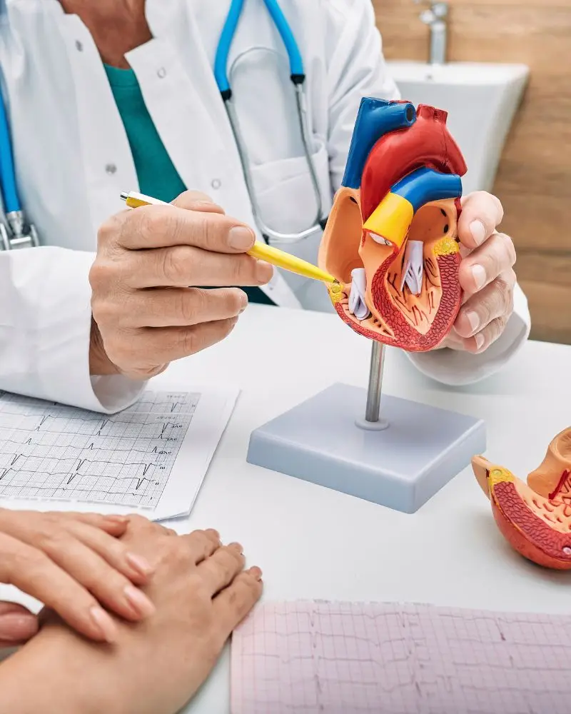



Nuclear imaging produces detailed information that only matters if it is interpreted well. Our cardiologists review your images against your symptoms and previous testing, and explain what they change.

General & Preventive Cardiology

Preventive & Non-Invasive Cardiology

Interventional & Vascular Cardiology
Treatment plans tailored to your symptoms, history, and individual needs.
Preventive, diagnostic, interventional, and vascular care in one practice.
Modern diagnostics for heart rhythm, function, structure, and blood flow.
Serving Pearland with additional locations expanding across Greater Houston.
Experienced physicians with advanced cardiovascular training and expertise.
We work with many leading insurance plans for convenient access to care.
Alliance Heart & Vascular is expanding to serve patients across Pearland, Greater Houston and surrounding communities.
Comprehensive cardiology, preventive cardiovascular care, diagnostic evaluation, and follow-up.
View Pearland LocationAlliance Heart & Vascular is expanding across Greater Houston and nearby communities, bringing convenient access to trusted cardiovascular care closer to where our patients live and work.
Alliance Heart & Vascular works with many major health insurance plans. Insurance participation can vary by plan, network, employer, and location.
Please call (346) 614-0002 before your appointment to verify your specific insurance benefits and network participation. Our team can help answer questions about scheduling, referrals, and insurance verification.
Nuclear testing is chosen when a standard stress test would be unreliable or has already left the question open. Common reasons include:
Nuclear imaging is particularly useful for deciding whether a narrowing already seen on another test is actually limiting blood flow, which is what determines whether treating it will help.
Nuclear stress testing helps investigate several cardiovascular conditions. Learn more about:
Nuclear testing is a scheduled investigation. If you have chest pain at rest, severe breathlessness, or fainting, call 911 immediately.
Do not wait for an office appointment.
Online appointment requests should never be used for potentially life-threatening symptoms.
Comparing rest and stress images distinguishes between muscle that is short of blood and muscle that has already been damaged:
The distinction between reversible ischemia and fixed scar is the central finding. Reversible ischemia means living muscle that could benefit from restored blood flow; scar will not improve with a stent.
A nuclear study takes longer than a treadmill test because imaging is performed twice, with a gap between the two sets of pictures.
A small cannula is placed and a low dose of radioactive tracer is given. The tracer travels to the heart muscle in proportion to blood flow.
A camera records how the tracer has distributed while you are at rest, establishing the baseline picture of blood supply.
You exercise on a treadmill, or receive medication that produces a similar effect, and a second dose of tracer is given at peak stress.
A second set of images is taken after the stress phase, showing blood distribution when demand on the heart was highest.
The two sets are compared. Areas that look normal at rest but poor under stress indicate reversible ischemia.
The study also produces an ejection fraction and wall motion assessment, adding functional information to the perfusion findings.
Preparation is important, particularly around caffeine, which interferes directly with the medication used in pharmacological studies:
Caffeine blocks the action of the medication used in pharmacological stress testing and can invalidate the entire study. Decaffeinated coffee and chocolate both count, so avoid them for the full 24 hours.
Nuclear imaging is aimed principally at coronary disease, and at deciding what should be done about it.
The central purpose. It shows whether a narrowing is genuinely limiting blood flow to the muscle it supplies. Read more about coronary artery disease.
Where exercise testing has been inconclusive, nuclear imaging clarifies whether exertional discomfort is ischemic. Read more about chest pain.
After a heart attack, imaging can show whether damaged muscle remains viable and might recover with restored blood flow. Read more about heart failure.
Breathlessness can be an angina equivalent, particularly in older patients and those with diabetes. Read more about shortness of breath.
Where risk is high but symptoms are ambiguous, imaging clarifies whether disease is already limiting flow. Read more about high cholesterol.
Not every patient needs every cardiac test. Testing should be selected based on your symptoms, examination, medical history, and cardiovascular risk. Depending on your individual situation, your cardiologist may recommend one or more of the following.
Measures the heart's electrical activity and may identify rhythm problems or other cardiac changes.
Uses ultrasound to evaluate heart function, chambers, valves, muscle, and structure.
Shows how the heart responds to physical activity and may help evaluate exercise-related symptoms.
Uses cardiac imaging during stress testing to assess blood flow to the heart muscle.
Records heart rhythm over time to help detect intermittent arrhythmias or palpitations.
Blood tests may evaluate cholesterol, blood sugar, kidney function, and other cardiovascular risk factors.
Certain patients may require additional cardiac imaging or coronary evaluation depending on their symptoms and risk profile. Your cardiologist will explain why a particular test is being recommended and how the results may influence your treatment plan.
The images are interpreted alongside your symptoms, risk factors, and any previous testing. What follows depends on the extent and severity of any abnormality:
A small area of mild ischemia is usually managed with medication. A large or severe area is what prompts angiography, because that is where restoring blood flow changes outcome rather than just symptoms.
Nuclear studies are often arranged after another test has raised a question without answering it — an inconclusive treadmill test, an abnormal EKG that cannot be interpreted, or a calcium score that needs putting in context.
A nuclear study is appropriate if you have:
Talk to our office — Monday to Friday, 8:00 a.m. to 4:30 p.m.
Call (346) 614-0002When a treadmill test leaves the question open, imaging answers it.
Whether your exercise test was inconclusive, your resting EKG cannot be interpreted, or a known narrowing needs assessing for significance, we perform and interpret nuclear stress testing on site.
New patients are welcome.
Serving patients throughout Pearland, Greater Houston, and surrounding areas.